
ADA backed X ray intervals and safety: children 12–24 months; high risk adults 6–18 months; low risk adults 24–36 months. Learn what to ask to avoid...
ADA Backed 6–36 Month Intervals: How Often Dental X Rays Are Needed
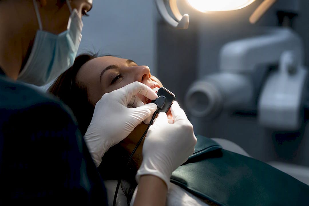
There is no one-size-fits-all schedule. X-rays are ordered based on your caries risk, age, and clinical findings, following the ADA’s 2026 guidance. For many low-risk adults, bitewing X-rays fall at intervals typically recommended in guidance, while higher-risk adults or kids in active development may need them more frequently, according to clinical judgment and ADA guidance.
TL;DR:
- The recommended frequency of dental X-rays depends on individual caries risk, age, and clinical findings, not a fixed schedule for everyone.
- High-risk patients, such as children with developing teeth or adults with active decay, often need closer monitoring every 6 to 24 months, while low-risk adults may only require imaging every 24 to 36 months.
- Digital imaging reduces radiation doses by 40 to 60 percent compared to film, and proper safeguards like collimation and shielding further minimize exposure.
- Panoramic and full-mouth surveys are only justified based on clinical need, not routine time intervals, and CBCT is reserved for complex cases due to higher exposure.
- Patients are advised to ask why each X-ray is necessary, share previous images, and consider alternatives to routine imaging to avoid unnecessary radiation exposure.
Table of Contents
- What Determines How Often You Need Dental X-Rays
- Typical Dental X-Ray Intervals by Patient Group
- Bitewing, Periapical, Panoramic, or CBCT: Which X-Ray Do You Actually Need?
- Is Dental X-Ray Radiation Safe? What the Numbers Actually Show
- How to Avoid Unnecessary Dental X-Rays
- How Clayton Dental Studio Applies These Guidelines in Practice
- Our Take: Why Blanket X-Ray Schedules Miss the Point
- Sources
What Determines How Often You Need Dental X-Rays
Your dentist isn’t flipping a coin or working off a calendar reminder. The single biggest factor is your caries risk, meaning how likely you are to develop new cavities based on your history, diet, and home care. The ADA is explicit that frequency should follow individual risk, not a fixed interval applied to everyone who walks through the door.
Several things feed into that risk picture:
- Your age and stage of dental development (a teenager with erupting molars needs different monitoring than a 60-year-old with stable teeth)
- Clinical signs your dentist sees during an exam, like visible decay, worn fillings, or gum recession
- Your medical history, including conditions or medications that raise cavity or bone-loss risk
- Whether you’re a new patient (baseline imaging), a recall patient (routine monitoring), or someone with a specific problem needing a focused image
New-patient visits almost always involve a first set of images so your dentist has a baseline to compare against later. After that, recall imaging gets spaced out or tightened based on what shows up. The 2026 ADA and American Academy of Oral and Maxillofacial Radiology guidance reinforces this: image only when an exam, your history, or prior films justify it, according to the ADA’s own release.
Typical Dental X-Ray Intervals by Patient Group
Here’s roughly what those risk-based intervals look like in practice, drawn from ADA guidance and how most dental practices apply it day to day.
- Children and teens with developing teeth: Bitewings are often taken every 12 to 24 months. Growing jaws and emerging permanent teeth mean more frequent monitoring is standard during mixed and transitional dentition.
- Adults with active decay or high caries risk: Expect bitewings every 6 to 18 months. Frequent cavities, deep fillings, or dry mouth from medication can all push your dentist toward closer monitoring.
- Low-risk adults with stable dental health: Every 24 to 36 months is typical. If your last several exams showed no new decay and your risk factors are low, there’s little clinical reason to image more often.
- Panoramic or full-mouth surveys: There is no fixed repeat schedule for panoramic or full-mouth surveys; decisions should be based on clinical need rather than a set interval. The ADA’s guidance on radiograph use makes clear these should be repeated only when clinical need justifies it, not because an insurance plan allows it every three or five years.
- New patients, treatment planning, or emergencies: These fall outside the routine schedule entirely. A cracked tooth, sudden swelling, or a first visit to a new dentist all call for imaging regardless of when your last X-ray was taken.
If your dentist wants to see you more often than these ranges suggest, ask why. A good answer will point to something specific, not a generic office policy.
Bitewing, Periapical, Panoramic, or CBCT: Which X-Ray Do You Actually Need?
Not every X-ray is the same, and the type your dentist orders says a lot about what they’re checking for.
- Bitewings capture the crowns of upper and lower back teeth biting together. These are the workhorse image for spotting cavities between teeth and are the main image tied to the interval ranges above.
- Periapical images show a single tooth from crown to root tip. Your dentist reaches for these when something specific is going on, like a suspected root infection, abscess, or a tooth that needs a root canal evaluated.
- Panoramic X-rays capture your entire jaw, sinuses, and all your teeth in one wide image. These help track wisdom teeth development, plan implants, or screen for broader issues like cysts or jaw abnormalities.
- Cone-beam CT (CBCT) produces a 3D image and delivers far more detail than a standard flat X-ray. Because of that added exposure, FDA guidance reserves CBCT for cases with a clear justified need, such as complex implant planning or evaluating a jaw fracture, not for routine checkups.
Is Dental X-Ray Radiation Safe? What the Numbers Actually Show
Some dental X-rays deliver less radiation than you’d absorb from a single day of ordinary background exposure from sunlight, soil, and the air around you. That comparison surprises a lot of patients who picture X-rays as a major radiation event. In dentistry, the dose is small, and the profession works hard to keep it that way.
Digital imaging can cut radiation dose by 40 to 60 percent compared to older film-based X-rays, according to the ADA. That single shift, from film to digital sensors, is one of the biggest safety upgrades dentistry has made in decades.
Behind that number sits a working principle called ALARA: As Low As Reasonably Achievable. In practice, ALARA means:
- Using the fastest digital receptors available to capture a diagnostic image with minimal exposure
- Applying rectangular collimation, which narrows the X-ray beam to the area actually being imaged instead of scattering radiation more broadly
- Limiting the number of images taken to only what’s clinically needed
- Using lead aprons and thyroid collars as a physical barrier during exposure
The FDA’s guidance for selecting radiographic patients backs all of this, and it’s the same framework that shapes how modern digital X-ray technology gets used in practice. If you’re pregnant or planning to become pregnant, tell your dentist. Dental X-rays are generally considered low risk with proper shielding, but your dentist may adjust timing or technique out of caution.
How to Avoid Unnecessary Dental X-Rays
You have more say in this than most patients realize. A few habits keep your imaging history efficient instead of redundant.
- Ask your previous dentist to transfer your existing X-rays before your first visit somewhere new, which is encouraged to improve patient communication and dental marketing efforts by attracting new dental patients. The ADA specifically encourages this to avoid duplicating images that already exist.
- Ask why whenever a new X-ray is recommended. A dentist following current guidance should be able to point to a specific exam finding, symptom, or time interval that justifies it.
- Bring up alternatives for monitoring, like a careful visual exam or an intraoral scan, when your risk is low and nothing suggests a hidden problem.
- Get a second opinion if a practice seems to image on every visit regardless of your history. That pattern doesn’t match how risk-based imaging is supposed to work.
Pro Tip: Keep a simple log of when and where you’ve had dental X-rays taken. Handing a new dentist that timeline speeds up their risk assessment and gives them one less reason to reorder images you already have.
How Clayton Dental Studio Applies These Guidelines in Practice
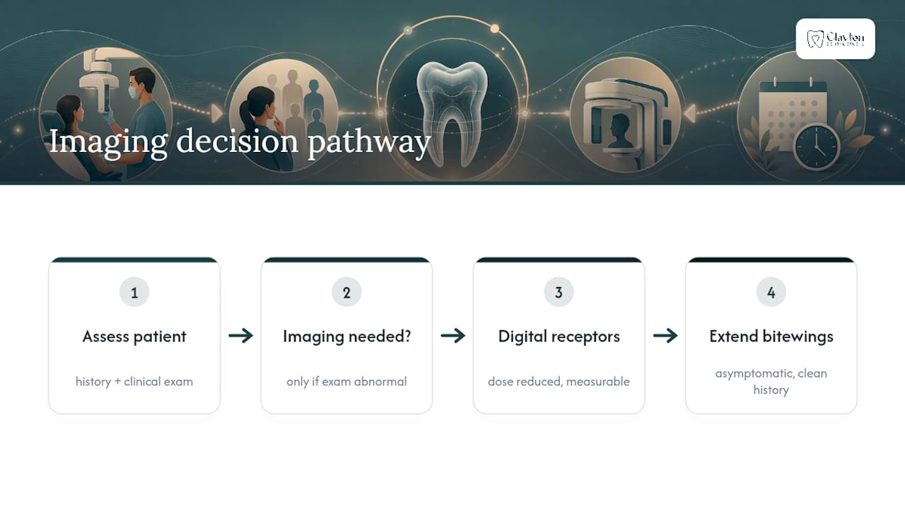
I’m Dr. Navneet Kamboj, and I’ve spent nearly a decade treating patients in the area, with a Doctor of Dental Surgery degree and additional restorative training. At Clayton Dental Studio, we start every imaging decision with a clinical exam and a look at your history first, not a routine we run on autopilot.
We rely on digital receptors because the dose reduction is real and measurable. For a routine recall patient with no new symptoms and a clean history, that often means bitewings stay on a longer interval rather than getting reordered out of habit. When something in the exam doesn’t add up, that’s when imaging earns its place.
Our Take: Why Blanket X-Ray Schedules Miss the Point
The 2026 ADA and AAOMR guidance settles something that’s been fuzzy for years: there is no default calendar for dental X-rays, and there never should have been one. The old habit of “X-rays every year” made scheduling simple for offices, but it never matched how cavities and bone loss actually behave in different people.
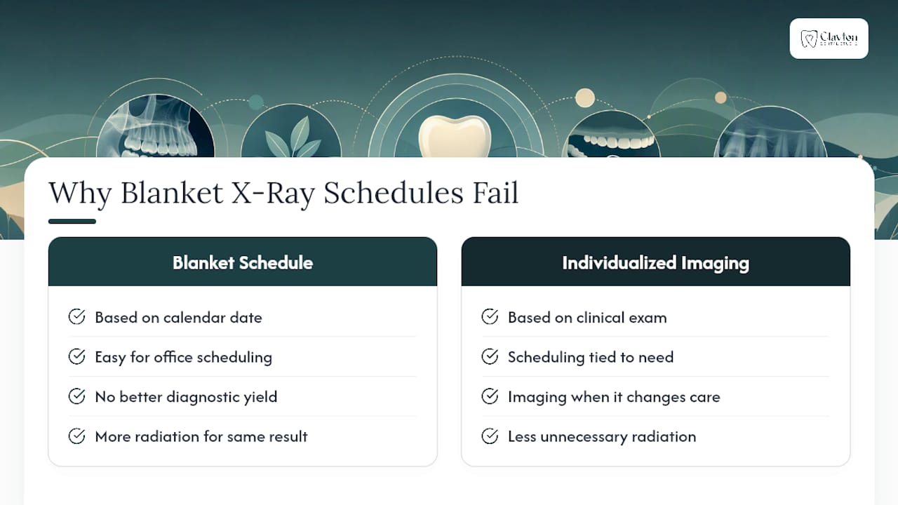
What conventional advice gets wrong is treating imaging frequency as a fixed benefit. More X-rays don’t mean better care. A low-risk adult imaged every year isn’t safer than one imaged every three, they’re just absorbing more radiation for the same diagnostic yield, which is exactly the pattern the ADA’s Dr. Erika Benavides pushed back on when framing dental imaging the way other medical imaging already works: ordered because an exam calls for it, not because a date came up.
If you take one thing from this, prioritize asking your dentist what specifically prompted a new X-ray order. That single question does more to protect you from unnecessary exposure than memorizing interval numbers ever will. Ready to get a clear, individualized read on your own risk and imaging needs? Book a visit with Clayton Dental Studio and get an answer that’s about you, not a chart.
— Clayton Dental Studio
This article is general information, not a substitute for advice from a qualified doctor. Consult a qualified healthcare professional about your own circumstances before acting on anything here.
Sources
- New Recommendations Confirm Dental X-Rays Most Effectively Used in Moderation | American Dental Association
- Selection of Patients for Dental Radiographic Examinations (FDA guidance)
- X-rays | MouthHealthy (ADA)
Recommended
Ready to Schedule Your Appointment?
Dr. Kamboj and the Clayton Dental Studio team are here to help. Call us or book online today.