Discover effective peri implantitis treatment options to save your implant. Learn step-by-step care for lasting dental health.
Peri-Implantitis Treatment: Your Complete Care Guide
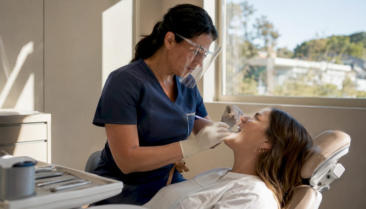
If you’ve been diagnosed with peri-implantitis, you’re likely wondering what comes next and whether your implant can be saved. The short answer is yes, in most cases, but treatment requires a structured, stepwise approach rather than a single quick fix. Peri-implantitis is a bacterial infection that causes inflammation in the gum tissue and progressive bone loss around a dental implant. It differs from peri-implant mucositis, which involves only soft tissue inflammation with no bone loss. Think of mucositis as the earlier, more reversible warning sign. Peri-implantitis is what happens when that warning goes unaddressed.
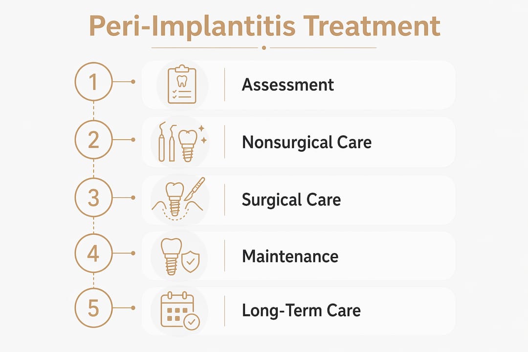
Here’s what effective management looks like at a glance:
- Start with nonsurgical treatment: Professional mechanical and chemical cleaning to remove bacteria and irritants around the implant.
- Use antibiotics selectively: Systemic antibiotics like metronidazole may be added to improve outcomes, but they’re not a standalone solution.
- Escalate to surgery when needed: If nonsurgical treatment doesn’t stabilize the infection, surgical access, resective, or regenerative procedures become necessary.
- Target measurable goals: Clinicians aim for pocket depths at or below 5 mm and minimal bleeding on probing.
- Commit to maintenance: Regular professional cleanings and monitoring are non-negotiable for long-term implant survival.
- Expect a management cycle, not a one-time cure: Peri-implantitis rarely resolves after a single round of treatment. Stabilization and ongoing monitoring are the realistic goals.
Table of Contents
- How peri-implantitis develops and what it looks like
- Nonsurgical peri-implantitis treatment: where care begins
- Surgical options when nonsurgical care isn’t enough
- Managing expectations and planning for long-term care
- Dr. Kamboj’s clinical perspective on treating peri-implantitis
- Peri-implantitis care at Claytondentalstudio in Humble, TX
- Key Takeaways
How peri-implantitis develops and what it looks like
Peri-implantitis doesn’t appear overnight. It typically begins as peri-implant mucositis, where bacteria accumulate around the implant and trigger localized gum inflammation. If that inflammation isn’t resolved, the infection spreads deeper, and the bone supporting the implant begins to break down. That bone loss is what separates peri-implantitis from mucositis and what makes it a more serious clinical concern.
Common symptoms you might notice include redness or swelling around the implant, bleeding when you brush or floss near it, a bad taste or odor that doesn’t go away, and in more advanced cases, visible bone loss on an X-ray or even implant mobility. Some patients feel pain or pressure around the site; others notice nothing at all until a routine exam catches it. That’s part of what makes early detection so valuable.
Clinically, your dentist will use a periodontal probe to measure pocket depths around the implant. Healthy implants typically show shallow, stable pockets. Deeper pockets, especially when they bleed on probing, signal active disease. Radiographic imaging, including cone beam computed tomography (CBCT), gives a three-dimensional view of the bone architecture around the implant and helps map the extent of bone loss before any treatment begins. CBCT imaging is particularly useful for surgical planning because it reveals the exact shape and depth of bone defects that a standard X-ray might underrepresent.
One detail worth understanding: peri-implantitis progresses more rapidly than periodontitis around natural teeth. The implant surface, unlike a natural tooth root, lacks a periodontal ligament to act as a biological buffer. That means the infection can advance faster and with less warning, which is exactly why regular monitoring matters so much once you have implants.
Nonsurgical peri-implantitis treatment: where care begins
The first line of peri-implantitis treatment is always nonsurgical. The goal at this stage is to disrupt and remove the bacterial biofilm that’s driving the infection, reduce inflammation, and create conditions where the tissue can begin to heal. This is done through a combination of mechanical and chemical debridement, sometimes supported by antibiotics.
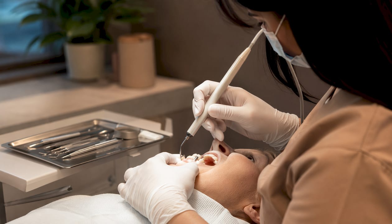
Mechanical debridement means physically removing plaque, calculus, and bacterial deposits from the implant surface and surrounding tissues. Your clinician will use instruments designed specifically for implant surfaces, typically titanium or carbon fiber curettes, ultrasonic devices, or air-polishing systems, to avoid scratching the implant while still cleaning it thoroughly. One step that significantly improves access is removing the implant-supported prosthesis before treatment. Prosthesis removal allows the clinician to reach areas that are otherwise hidden beneath the crown or bridge, enabling far more thorough decontamination.
Chemical debridement works alongside mechanical cleaning. Agents commonly used include:
- Chlorhexidine: An antiseptic rinse or gel applied directly to the pocket to reduce bacterial load.
- Citric acid and EDTA: Used to condition the implant surface and remove the biofilm layer that bacteria cling to.
- Hydrogen peroxide: Sometimes used as an irrigant to flush debris and bacteria from deeper pockets.
One thing to be clear about: mouthwash alone cannot treat peri-implantitis. Over-the-counter rinses don’t reach the bacteria living beneath the gumline, and they can’t remove calcified deposits. Professional intervention is the only way to address what’s actually driving the disease.
Antibiotics can play a supporting role. Systemic antibiotics combined with nonsurgical debridement improve probing depth reduction and treatment outcomes, but they must be used carefully. Antibiotic resistance is a real concern, and antibiotics should never substitute for thorough mechanical cleaning. Metronidazole is one of the most commonly used options, often in combination with amoxicillin, prescribed for a short course alongside active debridement.
Adjunctive therapies like lasers and locally delivered antimicrobials are sometimes offered as add-ons. The evidence here is more modest. Lasers and local antimicrobials show minimal additional clinical benefit when added to thorough mechanical debridement, so they shouldn’t be viewed as replacements for the fundamentals.
A critical step that’s often overlooked: removing plaque-retentive factors. Residual cement left behind from crown placement is one of the most common drivers of recurring inflammation. If excess cement is present around the implant, it must be removed before any other treatment can succeed. The same applies to poorly fitting prosthetic components or rough implant surfaces that trap bacteria.
Nonsurgical treatment is often enough for early-stage disease. If pocket depths reduce to a shallow and healthy range and bleeding on probing decreases significantly after a re-evaluation period, the condition may be considered stabilized. When those targets aren’t met, surgery becomes the appropriate next step.
Surgical options when nonsurgical care isn’t enough
Surgery for peri-implantitis isn’t a sign that treatment has failed. It’s a planned escalation that gives the clinician direct visual access to the implant surface and the surrounding bone defect. Surgical options fall into three main categories, and the right choice depends on the shape and severity of the bone defect.
Access surgery is the most conservative surgical approach. The gum tissue is reflected to expose the implant surface and the bone defect, allowing for thorough decontamination under direct vision. No bone is removed or added. This approach works best when the bone defect is relatively shallow and the primary goal is cleaning what couldn’t be reached nonsurgically.
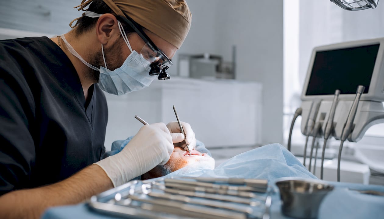
Resective surgery goes a step further. It involves reshaping the bone around the implant to eliminate pockets and reduce the areas where bacteria can accumulate. A technique called implantoplasty is often performed alongside resective surgery. In implantoplasty, the rough threaded portion of the implant that’s been exposed by bone loss is mechanically smoothed and polished. A smoother surface is harder for bacteria to colonize, which reduces the risk of reinfection. Resective approaches work best when the bone defect is horizontal and esthetic concerns are less of a priority, since the procedure can result in more implant surface being visible.
Reconstructive (regenerative) surgery takes the opposite approach: instead of removing bone, it attempts to rebuild it. This involves placing bone grafts, barrier membranes, or a combination of both to encourage new bone growth around the implant. Regenerative procedures are best suited for vertical or circumferential bone defects, where the geometry of the defect supports the graft material staying in place. The evidence for regenerative surgery is encouraging, though outcomes vary depending on defect morphology, implant surface characteristics, and how thoroughly the surface was decontaminated during the procedure.
Key considerations when choosing between these approaches:
- Defect shape: Vertical, contained defects favor regeneration. Horizontal, flat defects favor resection.
- Esthetic zone: Implants in visible areas require more careful planning to avoid recession that would expose the implant.
- Surface decontamination: Regardless of which surgical approach is chosen, thorough cleaning of the implant surface during surgery is the single most important factor in long-term success.
- Patient factors: Smoking, uncontrolled diabetes, and poor oral hygiene all reduce surgical success rates and must be addressed before proceeding.
Managing expectations and planning for long-term care
Peri-implantitis treatment is not a one-and-done procedure. That’s not a discouraging statement; it’s an honest one that helps you plan. The disease can be controlled and stabilized, but it requires ongoing commitment from both you and your dental team.
Prognosis varies. Some implants respond well to nonsurgical treatment and remain stable for years with proper maintenance. Others require surgery, and even after surgery, a small percentage of implants may eventually need to be removed if the infection cannot be controlled. The factors that most strongly predict a good outcome include early diagnosis, thorough treatment, and consistent maintenance.
Maintenance intervals matter. After active treatment, many patients with a history of peri-implantitis require more frequent professional cleanings than standard schedules. Your recall interval should be individualized based on your risk profile, including your history of periodontal disease, smoking status, diabetes control, and how well you maintain oral hygiene at home.
Soft tissue quality plays a bigger role than most patients expect. The health and thickness of the keratinized gum tissue around your implant significantly affects bone loss prevention and long-term stability. When there isn’t enough keratinized tissue, soft tissue grafting may be recommended as part of the overall treatment plan to create a more stable environment around the implant.
At-home care recommendations after treatment include:
- Brushing twice daily with a soft-bristle toothbrush, paying close attention to the gumline around the implant.
- Using interdental brushes or water flossers to clean between the implant and adjacent teeth.
- Avoiding tobacco, which significantly impairs healing and increases recurrence risk.
- Attending every scheduled maintenance appointment, even when the implant feels fine.
Recurrence is possible. Even after successful treatment, peri-implantitis can return if bacterial plaque is allowed to accumulate again. Monitoring at each maintenance visit includes probing, bleeding assessment, and periodic radiographs to catch any early signs of returning bone loss before they become serious.
Dr. Kamboj’s clinical perspective on treating peri-implantitis
I’m Dr. Navneet Kamboj, and over nearly a decade of treating patients in the Humble area, I’ve seen peri-implantitis at every stage, from early mucositis caught at a routine cleaning to advanced bone loss that required surgical intervention. What I’ve learned is that the patients who do best are the ones who understand what they’re dealing with and stay engaged in their care.
One of the most practical things I do before any peri-implantitis treatment is remove the prosthesis. It sounds simple, but it changes everything. You simply cannot clean an implant surface properly when the crown is in the way. Once we have direct access, we can see exactly where the bacterial deposits are, where the bone has been lost, and what kind of defect we’re working with. That information shapes the entire treatment plan.
I also spend time identifying plaque-retentive factors before we begin. Excess cement is the most common culprit I find, and it’s often the reason a patient’s implant has been struggling despite good home care. Removing it isn’t glamorous, but it’s frequently the single most impactful step we take.
When it comes to patient expectations, I’m always honest. I tell patients that we may need more than one round of treatment, that surgery is sometimes the right answer, and that their daily habits at home are just as important as what we do in the chair. That honesty builds trust, and it leads to better outcomes because patients who understand the process are more likely to follow through.
Peri-implantitis care at Claytondentalstudio in Humble, TX
If you’ve been diagnosed with peri-implantitis and you’re looking for a dental team that takes implant health seriously, Claytondentalstudio offers the kind of thorough, individualized care this condition requires. Dr. Kamboj combines nearly a decade of clinical experience with advanced diagnostic tools, including CBCT imaging, to assess bone defects accurately and plan treatment that fits your specific situation.
What sets the approach at Claytondentalstudio apart is the emphasis on getting the details right: removing the prosthesis for proper access, identifying and eliminating plaque-retentive factors, and building a maintenance schedule that keeps the disease from returning. Whether your case calls for nonsurgical debridement or a surgical intervention, the goal is always the same: stabilize the infection, protect the bone, and give your implant the best possible chance of lasting. You can explore the full range of dental implant services available at the practice, or reach out directly to schedule a consultation. The sooner peri-implantitis is addressed, the more options you have.
Key Takeaways
Effective peri-implantitis management requires a staged protocol combining mechanical debridement, selective antibiotics, defect-specific surgery, and a structured maintenance program to stabilize infection and preserve the implant long term.
| Point | Details |
|---|---|
| Nonsurgical treatment comes first | Mechanical and chemical debridement, with selective antibiotic support, is the starting point for all peri-implantitis cases. |
| Surgery is defect-specific | Access, resective, and regenerative surgery are chosen based on bone defect shape, severity, and esthetic considerations. |
| Pocket depth target is ≤5 mm | Clinicians aim for pocket depths at or below 5 mm with minimal bleeding on probing as the benchmark for disease stabilization. |
| Maintenance prevents recurrence | Professional cleanings at individualized intervals and consistent home care are required after treatment to prevent the disease from returning |
| Claytondentalstudio offers structured implant care | Dr. Kamboj provides individualized peri-implantitis treatment in Humble, TX, from diagnosis through long-term maintenance. |
Recommended
Ready to Schedule Your Appointment?
Dr. Kamboj and the Clayton Dental Studio team are here to help. Call us or book online today.