Learn what to expect from a dental bone graft procedure. Understand healing times and how it prepares your jaw for implants.
Dental Bone Graft: What to Expect Before Implants
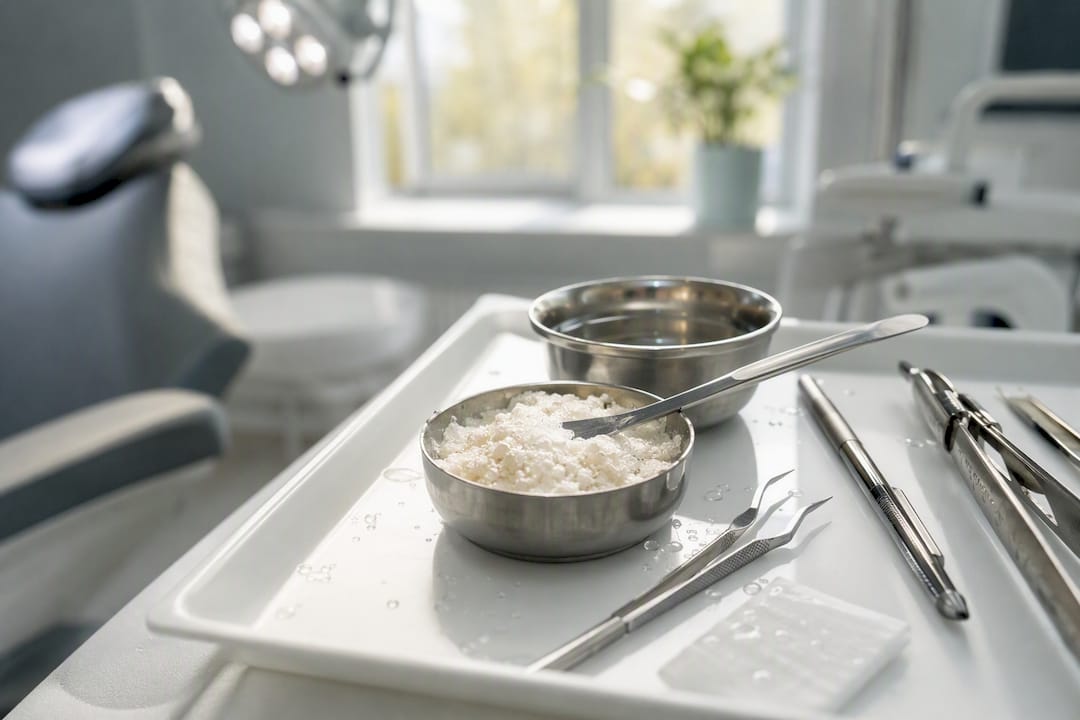
A dental bone graft rebuilds jawbone that has thinned or resorbed, usually to create a stable foundation for a dental implant. It works like scaffolding: the graft material holds space and signals your body to grow new bone into it, rather than replacing bone instantly.
Soft tissue over the graft site closes in about 1 to 2 weeks, but the bone itself needs far longer. Full integration typically takes 4 to 6 months for standard grafts, and up to 9 to 12 months for larger reconstructions. That gap between “it looks healed” and “it’s actually ready” is where most patients get impatient, and where timing mistakes happen.
- Definition: A graft replaces or builds up missing jawbone, most often before implant placement.
- Soft-tissue healing: 1 to 2 weeks.
- Bone integration: 4 to 12+ months, depending on graft size and type.
- Next step: If you’ve had a tooth missing for a while, wear dentures, or your dentist mentioned “not enough bone,” ask for a CBCT scan and a consult before assuming implants are off the table.
Quick fact: Most dental bone grafts succeed, but the timeline, not the surgery itself, is usually what determines whether an implant holds long term.
Key Takeaways
A dental bone graft rebuilds jawbone using a scaffold material, and success depends less on the surgery itself than on respecting the 4 to 12 month integration timeline before implant placement.
| Point | Details |
|---|---|
| Two-phase healing | Soft tissue closes in 1 to 2 weeks; bone integration takes 4 to 12+ months depending on graft size. |
| Material choice matters | Autograft offers the strongest biology; allografts, xenografts, and synthetics avoid a second surgery site. |
| Combining materials is common | Clinicians often blend a faster allograft with a slower-resorbing xenograft for volume and speed. |
| Imaging comes first | A CBCT scan, not a flat X-ray, should guide any grafting decision. |
| Plan with Clayton Dental Studio | CBCT-based evaluation and a clear, personalized implant timeline start with a single consultation. |
Table of Contents
- What Is a Dental Bone Graft, and Why Would You Need One?
- What Types of Bone Graft Materials Are Used?
- What Happens During the Bone Graft Procedure?
- Sinus Lift, Socket Preservation, or Ridge Augmentation: Which Applies to You?
- What Are the Risks, and How Successful Is Bone Grafting?
- How Long Does Recovery Take, and What Should You Do During It?
- How Much Does a Dental Bone Graft Cost, and Will Insurance Cover It?
- How Do You Choose the Right Dentist for a Bone Graft?
- How Does a Modern Dental Practice Approach Bone Grafting?
- Why Planning and Timing Matter More Than the Surgery Itself
- Ready to Find Out if You Need a Bone Graft?
- Frequently Asked Questions
- Sources
What Is a Dental Bone Graft, and Why Would You Need One?
Your jawbone needs stimulation to maintain its density, and chewing forces on natural tooth roots provide that stimulation. Pull a tooth, and the bone underneath starts resorbing within months. That’s the core problem grafting solves: it rebuilds the volume and density an implant needs to anchor securely, or it repairs bone lost to gum disease or injury.
Dentists recommend grafting for several distinct situations, and knowing which one applies to you changes what to expect:
- Implant site preparation — building up bone before or during implant placement when the ridge has thinned.
- Socket preservation — grafting immediately after an extraction to stop the natural collapse of the socket walls.
- Ridge augmentation — widening or heightening a jaw ridge that has resorbed over years of missing teeth.
- Sinus lift — adding bone beneath the sinus floor in the upper jaw, where sinus position often limits implant depth.
- Periodontal defect repair — filling bone pockets created by advanced gum disease.
- Trauma repair — reconstructing bone lost to injury or fracture.
A graft is not a bone transplant in the sense of a swap. It’s a scaffold your own cells colonize and remodel, which is why the material’s biological behavior matters as much as its shape.
Before any of this happens, your dentist needs to see what’s actually there. A 3D CBCT scan measures bone height, width, and density in ways a flat X-ray simply can’t, and it’s now standard practice for planning graft and implant cases together. Clayton Dental Studio uses CBCT imaging for exactly this reason: guessing at bone volume leads to surprises mid-surgery.
Pro Tip: If a clinic recommends grafting without ever taking a 3D scan, ask why. A panoramic X-ray alone can’t reliably measure ridge width, and width is often the deciding factor in whether you need a graft at all.
What Types of Bone Graft Materials Are Used?
The material choice affects healing speed, long-term stability, and cost, and it’s one of the first decisions you and your dentist will make together.
Autograft (your own bone, typically harvested from the jaw, chin, or hip) is the biological gold standard. It carries living cells and growth signals no other material fully replicates, so it integrates predictably. The tradeoff is a second surgical site, which means more discomfort and a longer overall procedure. Most implant-prep cases don’t need it.
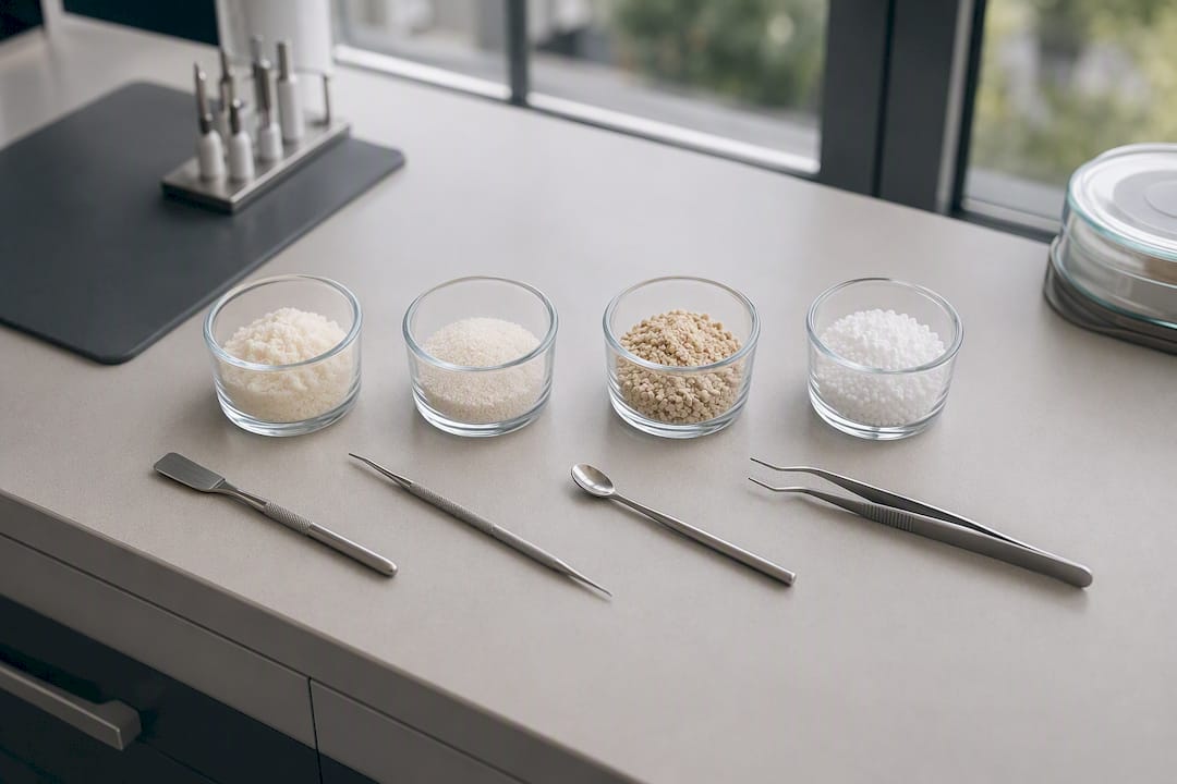
Allograft (processed donor bone from a tissue bank) skips the second surgery site and remains widely used for moderate defects. It resorbs and remodels faster than some alternatives, which suits cases where you want the graft site ready sooner.
Xenograft (commonly bovine-derived, or DBBM) resorbs much more slowly. That slow turnover is a feature, not a flaw, when the goal is long-term volume maintenance rather than fast remodeling, such as under a sinus lift where the graft needs to hold its shape for years.
Synthetic/alloplastic materials (hydroxyapatite, beta-tricalcium phosphate) are lab-made, carry no disease-transmission risk, and are available in unlimited supply, though they lack the biological signaling of living bone.
| Material | Source | Resorption speed | Best suited for |
|---|---|---|---|
| Autograft | Patient’s own bone | Moderate | Large defects, strongest biology |
| Allograft | Donor bone bank | Faster | Moderate defects, socket preservation |
| Xenograft | Animal-derived (often bovine) | Slow | Sinus lifts, long-term volume needs |
| Synthetic | Lab-manufactured | Varies by formula | Smaller defects, availability-driven cases |
| Factor | Autograft | Allograft/Xenograft/Synthetic |
|---|---|---|
| Donor-site morbidity | Yes, second surgical site | None |
| Supply limits | Limited by patient anatomy | Essentially unlimited |
| Biological signaling | Strongest (osteogenic) | Osteoconductive/inductive only |
| Typical use | Complex reconstructions | Routine implant prep |
Clinicians frequently blend materials rather than pick one. Combining a faster-resorbing allograft with a volume-stable xenograft, often close to a 1:1 ratio, is a common approach for sinus augmentation because it balances quick regeneration with lasting bulk.
Pro Tip: Ask your dentist which material they’re using and why. “We use allograft because it remodels quickly for your socket size” is a very different answer from “we use xenograft because your sinus lift needs to hold volume for years,” and both are legitimate, defect-specific reasoning.
What Happens During the Bone Graft Procedure?
Before scheduling surgery, your dentist reviews your medical history, current medications, and smoking status, since nicotine measurably slows healing and raises graft failure risk. The CBCT scan taken during evaluation maps exact bone dimensions, and any active infection at the site gets treated first. Skipping this step is one of the more common reasons grafts fail.
The procedure itself typically follows this sequence:
- Anesthesia or sedation — local anesthesia handles most graft cases; larger reconstructions may use IV sedation or, rarely, general anesthesia.
- Incision and access — the dentist opens a small flap of gum tissue to expose the bone.
- Graft placement — the chosen material is packed into the defect or deficient area.
- Membrane and fixation — a collagen membrane often covers the graft to keep soft tissue from invading the space, sometimes secured with tiny pins or screws.
- Closure — the gum tissue is sutured back over the site.
Immediately afterward, expect swelling that peaks around day 2 or 3, manageable pain typically controlled with over-the-counter or short-course prescription medication, and possibly minor bleeding or oozing for the first 24 hours. Your dentist will give you a short list of restrictions:
- No smoking, which measurably slows healing.
- Soft foods only for several days.
- No vigorous rinsing or spitting, which can dislodge the clot.
- Ice on the outside of the face during the first 48 hours to control swelling.
If nonresorbable membranes or fixation screws were used, a follow-up visit a few weeks later removes them; resorbable materials dissolve on their own.
Pro Tip: Fill your pain prescription before surgery, not after. Swelling and discomfort usually build over the first 48 hours, and you don’t want to be driving to a pharmacy while your face is swollen.

Quick fact: Most dental bone graft procedures are outpatient, done under local anesthesia, and completed in under an hour for a single site.
Sinus Lift, Socket Preservation, or Ridge Augmentation: Which Applies to You?
The right procedural approach depends entirely on where and why bone was lost.
- Socket preservation happens right after an extraction, packing the empty socket with graft material before it has a chance to collapse inward. It’s the least invasive of the group and often pairs directly with a future implant.
- Ridge augmentation rebuilds a jaw ridge that’s already thinned or narrowed, usually from years of a missing tooth. It’s frequently done as its own procedure ahead of implant placement.
- Sinus lift raises the sinus floor in the upper back jaw to create room for an implant, since the maxillary sinus often sits too close to the ridge for adequate implant depth.
- Block grafts use a solid piece of bone (often autograft) for severe height or width loss that particle grafts can’t correct.
Timing varies by scenario. Minor defects sometimes allow simultaneous augmentation, meaning the graft and implant go in during the same visit. Larger defects require staged grafting: the graft heals first, typically around 6 months, before the implant goes in at all.
- Extraction with immediate socket preservation.
- Healing period (weeks to months, defect-dependent).
- Re-evaluation with imaging.
- Implant placement, either simultaneous with grafting (minor cases) or staged after full integration (major cases).
Large reconstructions involving multiple sites or significant bone volume sometimes require an oral surgery center with sedation capabilities beyond what a standard dental chair offers.
| Procedure | Typical trigger | Timing pattern |
|---|---|---|
| Socket preservation | Immediately after extraction | Often staged, 3 to 4 months before implant |
| Ridge augmentation | Long-standing missing tooth, narrow ridge | Staged, 4 to 6 months |
| Sinus lift | Insufficient bone height in upper jaw | Staged, 6 to 9 months |
| Block graft | Severe height/width loss | Staged, 6 to 12 months |
What Are the Risks, and How Successful Is Bone Grafting?
Most dental bone grafts succeed, and complications are uncommon when the site is properly evaluated and cared for. Still, no surgical procedure is risk-free, and knowing the warning signs helps you catch problems early.
- Infection at the graft site, usually preventable with proper hygiene and prescribed antibiotics when indicated.
- Graft failure or partial resorption, more common in smokers or patients with uncontrolled diabetes.
- Nerve irritation or numbness, rare, and typically temporary when it occurs near the lower jaw.
- Sinus complications, specific to sinus lift procedures, including membrane perforation.
- Bleeding, usually minor and self-limiting.
| Risk factor | Effect on outcomes |
|---|---|
| Smoking | Significantly slows healing, raises failure risk |
| Uncontrolled diabetes | Impairs bone regeneration and immune response |
| Poor oral hygiene | Increases infection risk at graft site |
| Untreated infection pre-op | Major contributor to graft failure |
Rushing implant placement before the bone has fully integrated is one of the most preventable causes of implant failure. Patience with the timeline matters as much as surgical skill.
Pro Tip: If your gums look fully healed at three weeks, that’s soft-tissue healing, not bone integration. Resist the urge to push your dentist for an early implant date. The bone underneath is still remodeling.
How Long Does Recovery Take, and What Should You Do During It?
Recovery happens in two very different phases, and confusing them causes most of the frustration people feel with this process.
Soft-tissue healing wraps up in 1 to 2 weeks: swelling fades, stitches dissolve or get removed, and the gum looks normal. Bone integration is a separate, slower process. Standard grafts need 4 to 6 months to vascularize and mature; larger reconstructions like sinus lifts or block grafts can take 9 to 12 months or longer before they’re ready to support an implant.
- Days 1 to 3: Ice, soft foods, prescribed pain management, avoid spitting or straws.
- Days 4 to 14: Transition to a slightly firmer soft diet, resume gentle brushing away from the site, follow-up visit if sutures need removal.
- Weeks 3 to 8: Gum tissue fully closed; resume normal hygiene routine.
- Months 2 to 12: Bone maturation continues quietly, with periodic imaging checks to confirm readiness for implant placement.
Watch for signs that warrant a call to your dentist rather than a wait-and-see approach:
- Fever above 101°F.
- Swelling that worsens after day 3 instead of improving.
- Pus or foul-tasting drainage from the site.
- Numbness that persists beyond a few days.
- Visible graft material or a sensation the site has “opened up.”
Once implants are in place, keeping the surrounding tissue healthy long-term matters just as much as the graft itself. Our guide to peri-implantitis prevention covers how to protect that investment for years, not just months.
Quick fact: Aftercare that avoids smoking and follows soft-diet guidance for several days measurably reduces infection risk at the graft site.
How Much Does a Dental Bone Graft Cost, and Will Insurance Cover It?
Cost depends on graft size, material choice, whether sedation beyond local anesthesia is needed, and how many follow-up visits the case requires. A small socket preservation graft costs far less than a sinus lift or block graft requiring specialized materials and a longer recovery-monitoring schedule.
Insurance coverage is inconsistent. Some plans classify grafting as a medically necessary restorative procedure and cover a portion; others treat it as a preparatory step for an elective implant and cover little or nothing. It genuinely depends on your specific plan’s language.
- Ask for a fully itemized estimate before treatment, not a rough range.
- Confirm whether your insurer requires pre-authorization for grafting.
- Ask about financing or phased treatment if the graft and implant together strain your budget.
Our dental implant cost breakdown walks through how graft and implant expenses typically stack together in a full treatment plan.
How Do You Choose the Right Dentist for a Bone Graft?
Not every general dentist performs grafting routinely, and the skill gap between an experienced provider and an occasional one shows up in outcomes.
- Confirm training and case volume in oral surgery or periodontics, specifically with graft and implant cases.
- Ask whether they use CBCT imaging as standard practice, not just for complex cases.
- Request before-and-after examples of similar procedures.
- Ask directly: what’s your expected timeline for my specific case, which graft material would you use and why, and what’s your complication rate?
Red flags worth walking away from:
- A provider who skips imaging and estimates bone volume by eye.
- Vague or rushed answers about consent and alternatives.
- No willingness to discuss material choices or timeline tradeoffs.
- Pressure to schedule the implant immediately after grafting, regardless of healing stage.
How Does a Modern Dental Practice Approach Bone Grafting?
A well-run bone grafting workflow starts with imaging, not assumptions. At Clayton Dental Studio, that means:
- CBCT imaging to map exact bone volume and density before any treatment plan is proposed.
- A shared conversation about graft material options, weighing your specific defect against recovery time and long-term goals.
- A clear, written timeline for when implant placement can realistically happen.
- Scheduled follow-up checks to confirm the graft is integrating as expected before moving forward.
The goal isn’t just placing a graft. It’s building a bone foundation that will hold an implant securely for decades, which means getting the imaging, material choice, and timing right the first time.
Why Planning and Timing Matter More Than the Surgery Itself
The surgical part of a bone graft is often the easiest part of the process. What determines whether it actually works is patience with the biology afterward. A grafted site is living tissue: it needs blood supply, cellular activity, and eventually mechanical stimulus from chewing to hold its density once an implant is placed. Rushing the sequence, or assuming healed gums mean healed bone, is where outcomes go sideways. Realistic timelines aren’t a formality. They’re the difference between an implant that lasts and one that doesn’t.
Ready to Find Out if You Need a Bone Graft?
If you’ve been told you don’t have “enough bone” for an implant, that’s usually the start of the conversation, not the end of it. Clayton Dental Studio offers in-house CBCT imaging, so instead of guessing at bone volume from a flat X-ray, you get an accurate 3D picture and a specific, honest timeline for your case.
What that first visit typically includes:
- A CBCT scan to measure your exact bone height, width, and density.
- A conversation about graft material options suited to your specific defect.
- A clear implant timeline, including whether staged or simultaneous grafting fits your case.
- Financing and insurance guidance so cost surprises don’t derail your treatment plan.
If you’re in the Humble area and ready to find out where you stand, schedule a consultation to get imaging done and a real plan built around your bone, not a generic estimate.
Frequently Asked Questions
What is a dental bone graft? A dental bone graft is a procedure that rebuilds or adds jawbone volume, most often to prepare a site for a dental implant or to repair bone lost after an extraction, gum disease, or injury.
How long does bone graft healing take before I can get an implant? Soft tissue heals in about 1 to 2 weeks, but the bone itself typically needs 4 to 6 months for standard grafts and 9 to 12 months for larger reconstructions before it’s ready to support an implant.
Does a dental bone graft hurt? Most patients report manageable discomfort similar to a tooth extraction, controlled with prescribed or over-the-counter pain medication. Swelling typically peaks around day 2 or 3 and improves steadily after that.
Is a bone graft always necessary before implants? No. If you have adequate bone height and width, you may go straight to implant placement. Grafting becomes necessary when imaging shows insufficient volume or density to support an implant securely.
What’s the difference between an allograft and a xenograft? An allograft comes from a human donor bone bank and tends to remodel faster; a xenograft, often bovine-derived, resorbs more slowly and is frequently chosen when long-term volume maintenance matters more than speed.
When should I call my dentist after a bone graft? Call if you develop a fever above 101°F, swelling that worsens after day 3, pus or foul drainage, persistent numbness, or a sensation that the graft site has reopened.
This article is general information, not a substitute for advice from a qualified doctor. Consult a qualified healthcare professional about your own circumstances before acting on anything here.
Sources
- Dental bone graft — Cleveland Clinic
- Bone grafting — Mount Sinai
- Bone Grafts in Dental Medicine: An Overview of Autografts, Allografts and Synthetic Materials — MDPI/PMC
- Optimized bone grafting — Periodontology 2000 (Ovid)
Recommended
Ready to Schedule Your Appointment?
Dr. Kamboj and the Clayton Dental Studio team are here to help. Call us or book online today.